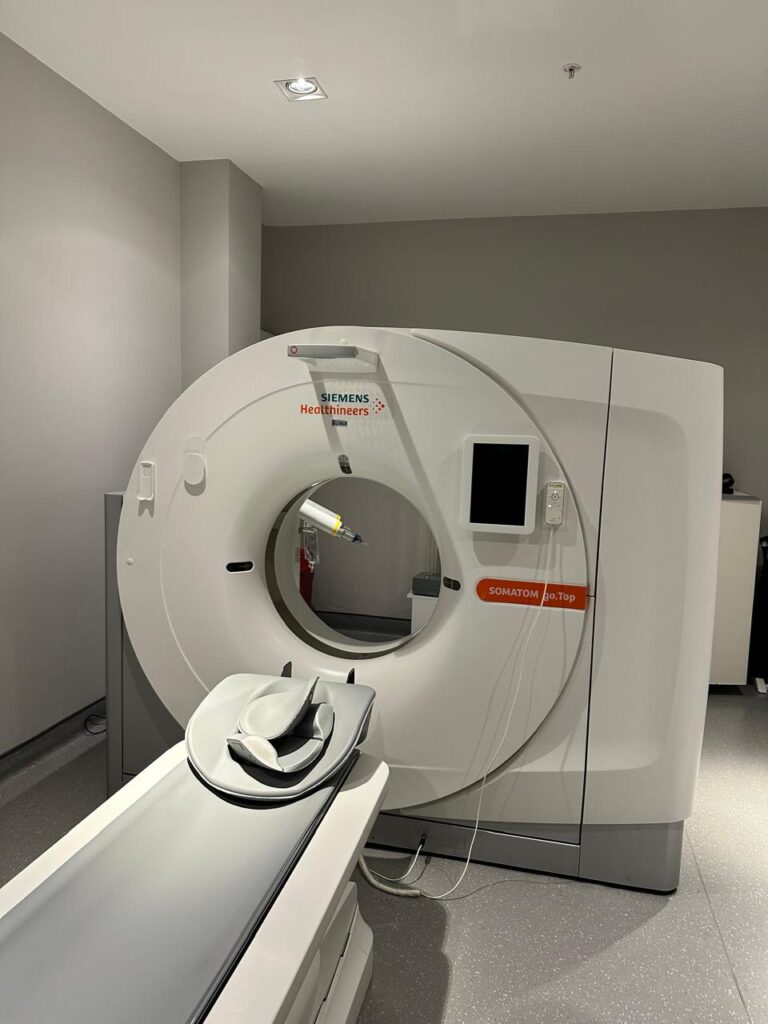
A CT scan, short for computed tomography scan, is a medical imaging technique that uses X-rays to produce detailed images of the inside of your body. Unlike traditional X-rays which provide a flat, 2D image, a CT scan creates cross-sectional images or "slices" of the body, offering a more comprehensive view.
A CT scanner employs an X-ray tube that rotates around the patient while detectors capture the X-ray images as they pass through the body. A powerful computer then processes this data to generate detailed images. These images can be viewed individually or combined to create a 3D representation.
Why Are CT Scans Used?
CT scans are versatile and used for a wide range of diagnostic purposes:
Injury assessment: Detecting fractures, internal bleeding, or soft tissue damage.
Cancer diagnosis and staging: Identifying tumors, determining cancer spread, and evaluating treatment response.
Cardiovascular evaluation: Assessing blood flow, detecting blockages, and evaluating heart conditions.
Pulmonary assessment: Diagnosing pneumonia, emphysema, or lung cancer.
Abdominal imaging: Investigating pain, inflammation, or organ abnormalities.
CT scans offer several advantages:
Detailed images: Provides clear visualization of various body structures.
Rapid results: Enables quick diagnosis and treatment planning.
Non-invasive: Generally safe and doesn't require surgical procedures.
Versatility: Can be used to examine almost any part of the body.
However, it's essential to consider the limitations:
Radiation exposure: While minimal, repeated CT scans can increase exposure.
Contrast dye allergies: Some individuals may experience allergic reactions to contrast dye.
Cost: CT scans can be more expensive compared to other imaging methods.


